Introduction
The dog is attractive species to discover genes underlying the homologous, relevant diseases in humans due to the high relevance of canine diseases to those in humans and the intrinsic importance of dogs to humans as special companions with shared environments. The domestic dog species is divided into over 300 pure breeding populations known as breeds. Many breeds are characterized by reduced genetic diversity related to small numbers of founders, popular sires whose allelic pool is over represented in subsequent generations, and changes in breed popularity over time (Quignon et al., 2007). Shared phenotypic characteristics of human and canine hip dysplasia (HD) are hip joint laxity accompanied by hip subluxation. Human HD, referred to as developmental dysplasia of the hip, occurs with a frequency ranging from 5, 13% (Zhou et al., 2010). Canine HD, a complex trait, is a major veterinary problem occurring with a frequency up to 75% in mixed and pure breed dogs of the approximately 70 million dogs in American households. Canine HD is a complex genetic skeletal disease observed in the majority of breeds of dogs and in almost all large-sized breeds of dogs. Canine HD is characterized by delayed onset of the capital femoral ossification, laxity of the joint, loss of congruency between acetabulum and femoral head, subluxation up to luxation, and often secondary osteoarthrotic changes. The first report of canine HD was reported in 1935. A study in 2003 showed that the prevalence of Canine HD was 19.3% in the general population of pet dogs. The percentage of dysplasia for these breeds in that study was 35.4% for Rottweiler’s, 32.9% for German Shepherds, 30.3% for Golden Retrievers and 27% for Labrador Retrievers (Nganvongpanit et al., 2008). The Labrador retriever demonstrates a 3.4-fold increase in risk for the development of HD (Goldberg and Rubin, 1989). Moreover, the majority of canine HD (80%) had osteoarthritis (OA). So far, the initiating factors are unknown, and the rate and extent of the development of HD are variable, but the risk factors are both genetic and environmental. As mentioned above, most of the dogs with HD in the above mentioned study also had OA, but the standardized diagnostic protocol consists of the clinical sign, a physical examination and evaluation of the radiographic results, which cannot detect OA in its early stages (Nganvongpanit et al., 2008).
Worldwide, the Orthopedic Foundation for Animals (OFA) has been scoring hip radiographs and releasing some of the records publicly over the last 40 years. In a previous study showed that a consistent genetic improvement has accumulated. The genetic improvement was limited by the fact that the selection criteria of the majority of the breeding dogs had low accuracy. Criteria for canine HD evaluated in scoring schemes include congruency of acetabulum and femoral head, the extent to which the femoral head is fully encompassed by the acetabulum, shallowness of the acetabulum and (sub-)luxation of the hip joint, and smoothness and radiographic sharpness of bony structures. In Europe, Fédération Cynologique Internationale (FCI) has made uniform as much as possible the scoring of hip joints in all registered breeds of dogs. Frequency and distribution of severity of scores vary largely between breeds, but clinical and radiographic signs are consistent across the different breeds of dogs (Marschall and Distl, 2007).
In the past, the uses of molecular genetics in commercial applications of marker-assisted selection have focused on the use of individual genes or a few quantitative trait loci (QTL) linked to markers. But until recently single-nucleotide polymorphism (SNP) genotyping technologies have provided for the first time the opportunity to rapidly screen large numbers of samples for genetic variation associated with increased risk for disease. SNP Microarray provides data on both genotype and signal intensity, the combination of which can be used to generate information on chromosomal segment copy number.
Previous study is canine HD in German shepherd dogs the using micro satellites (MS) marker. A whole genome scan for QTL was performed in German shephered dogs and 11 paternal half sib families, including a total of 459 purebreed German shepherd dogs with sires, dams, and offspring, were genotyped for 261 microsatellites. QTL for canine HD were located on nine different canine chromosomes: 1, 3, 4, 8, 9, 16, 19, 26, and 33. (Marschall and Distl, 2007).
We present data for the first time to demonstrate that canine HD is predictable from genomic data so that selection decisions. This implies that human HD could also be predicted and suitable preventativemanagement could be applied to identify susceptible individuals who may be missed by physical screening and ultrasound and reduce the prevalence hip OA by pre-emptive intervention.
Materials and Methods
Animal
Forty eight native Labrador Retriever dogs were categorized into two groups. Twenty four dogs were low risk hip dysplasia (HD) group consisting of 13 male and 11 female. Another group had 24 dogs which were high risk HD group consisting of 13 male and 11 female. Blood samples were collected for genomic DNA isolation using Wizard genomic DNA purification kit (Promega, USA) from 48 and analyzed concentration and purity at absorbance of 260 nm and 280 nm using ND-1000 spectrophotometer (Nanodrop, USA).
Phenotypic traits
Radiology has commonly been used to diagnose CHD. The technique has been standardized worldwide, although there is some variation in radiograph evaluation. There are three (somewhat different) international scoring methods: the FCI, the OFA, and the British Veterinary Association/Kennel Club methods. The FCI scoring method is used in most mainland European countries, Russia, South America, and Asia. The OFA approach is used exclusively in the USA and Canada, and the BVA/KC method is used in Britain, Ireland, Australia and New Zealand. We used the OFA method, OFA method divided 7group and each group is excellent (0 point), good (1~6point), fair (7~8 point), borderline (9~12), mile (13~18), moderate and severe (18~). This study radiograph score divided into two groups low risk, high risk was divided into. Low risk group average is 3.3 point; high risk group average is 15.3 point.
SNP Genotyping
Canine sample genotyped on the infinium canine SNP22 beadChip (Illumina Inc., San Diego, CA) with ~22,000 SNPs across he genome (http://www.illumina.com/documents/ products/datasheets/datasheet_canine_snp20.pdf). The majority (92.3 %) of the Illumina SNPs had completed calls. We also removed SNPs with minor allele frequency (MAF) below 1%. The final analysis contained 21,455 SNPs for the Illumina array, the mean and median MAF were 0.2589 and 0.2399, respectively.
Statistical Analysis
Genotypes were tested for Hardy-Weinberg equilibrium (HWE) to identify possible genotyping errors using the Chi-square test in the R/SNPassoc Package (R Development Core Team). SNPs with HWE (P <0.05), fail to call (> 80%), monomorphic SNPs and minor allele frequency (< 0.01) were removed in this QTL study. The association between a given SNP and the outcome was assessed using the R/SNPassoc package (http://cran.r-project.org/web/packages/SNPassoc/index.html) (model 1).
[model I] yS exSNPeijijn=+++ y : hip dysplasia phenotype μ : Full average Sexi : Genotype effect (i=Female, Male and Male Castration) SNPj : Genotype effect (j=AA, AB, BB) eij: Random error, N~(0, ve2)
Results
SNP analysis identified three different chromosomes for canine HD and each chromosome location (Fig. 1-2, Table 1.). Figure 1 and Table 1 show the plots summarize the genome-wide association results for hip dysplasia. The genome-wide P value (-log10 p) of the t test for the SNP effect are plotted against position on 4, 38, X chromosome and Locate on 4, 38, X chromosome SNPs position, t-statistical analysis information. Chromosome number is plotted in the x-axis. Horizontal line indicates the threshold P < 0.0001.
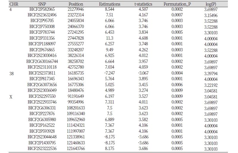
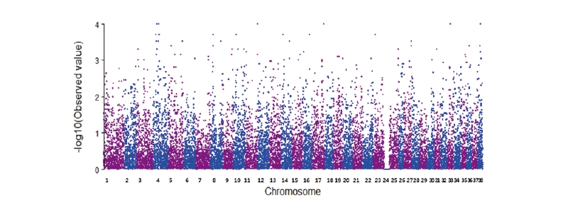
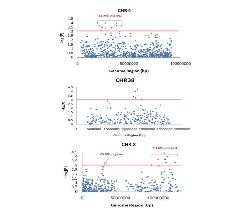
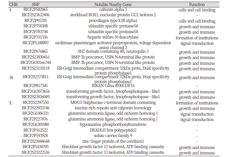
Table 2 shows gene function and notable nearby gene including significant SNPs. as shows notable nearby gene function is divided 4groups. Each group is cells and cell binding, growth and immune, signal transduction, formation of institutions. Notable nearby gene is caitenin alpha 3 (CTNNA3), stork head BOX1 (STOX1), nucleolar protein GU2 isoform 2 (DDX50), procollagen type XⅢ alpha1 (COL1A1), ubiauitin specific protease54 (USP54), heparin sulfate N-deacetylase (Ndst), urokinase plasminogen activator preproprotein (PLAU), voltage dependent anion channel 2 (VDAC2), SH2 domain containing 4B (Sh2d4b), neurequlin 3 (NRG1), BMP 3b precursor, USP6 N-terminal like protein (USP6NL), ER-Golgi intemediate compartment 32kDa prote, Dual specificity protein phosphatase1, RIKEN Cdna 4930513F16, Transforming growth factor (TGF) , lysophospholopase - like1, MOCO Sulphurase c-terminal domain containing (MOSC), leucine rich repeats and calponin homology (LRCH), glutamine ammonia ligase (GLUL), odd oz/tenm homolog 1 (ODZ1), hypoxantine phosphoribosyltransferse (HGPT), DEADLH box polypeptide2, solute carrier family 9 (SLC9A9), zinczinc finger protein of the cerellum3, fibroblast growth factor 13 isoform4 (FGF13 isoform4), ATP binding cassette (Abc) as notable nearby gene revealed a total of 27.
Discussion
We chose Labrador Retriever, as this breed represents the canine HD. From 1974 to 2009 provided by OFA hip dysplasia statistic Bulldog is NO1 in the population studied is 467 dogs and the hip dysplasia 73.2 percentage. According to research provided by OFA hip dysplasia the most many number of breed is Labrador Retriever, Golden Retriever, German Shepherd. Labrador Retrievers are especially studied the highest in 208,931. We performed hip dysplasia of Labrador Retriever SNP 22K chip. canine HD of Labrador Retriever discovered 25 SNPs (Table 1); 25 SNPs located on chromosome 4, 38, X. Until now, research on hip dysplasia SNPs were on chromosome 1, 3, 4, 8, 9, 11, 16, 19, 26, 30, 33 (Budsberg et al., 2006; Marschall and Distl, 2007). In this study, chromosomes 4, 38, X found in the significant SNPs, but a previous study published in the chromosomes of two hip dysplasia 1, 3, 4, 8, 9, 11, 16, 19, 26, 30, 33 overlap in chromosome 4, but in other 38, x chromosome and had found that these SNPs is significant. Hip dysplasia in Labrador Retriever 25 SNPs was found that a variety of roles (Table 2). Significant cell and cell binding, growth and immunity, formation of institutions, signal transduction, divided into four. Also found notable nearby gene is 27 (Table 2). Notable nearby gene is CTNNA3, STOX1, DDX50, COL1A1, USP54, Ndst, PLAU, VDAC2, Sh2d4b, NRG1, BMP 3b precursor, USP6NL, ER-Golgi intemediate compartment 32kDa prote, Dual specificity protein phosphatase1, RIKEN Cdna 4930513F16, TGF, lysophospholopase - like1, MOSC, LRCH, GLUL, ODZ1, HGPT, DEADLH box polypeptide2, SLC9A9, zinc finger protein of the cerellum 3, FGF13 isoform 4, Abc.
Until current research about the human and canine HD association gene is pregnancy associated plasma protein A2 (PAPP-A2) (Jia et al., 2012), cartilage oligomeric matrix protein (COMP) , matrilin 3 (MATN3) (Kim et al., 2011), collagen, type IX, alpha 1,2,3 (COL9A·1,2,3) (Dahlqvist et al., 2009; Kim et al., 2011; Nakashima et al., 2005), collagen, type II, alpha 1 (COL2A1) (Kannu et al., 2011), solute carrier family 26 (SLC26) (Hinrichs et al., 2010), fibrillin 2 (FBN2) (Friedenberg et al., 2011), transforming growth factor, beta 1 (TFGβ1), interleukin 6 (IL-6) (Kolundzic et al., 2011), interleukin beta 12 (IL-12β) (Zhou et al., 2010), aspirin (ASPN) (Shi et al., 2011), bone morphogenetic protein (BMP) (Baker-LePain and Lane, 2010), matrix metalloproteinase-1 (MMP-1) (Ray et al., 2005).
These genetic functions of PAPP-A2 is cell differentiation, regulation of cell growth, COMP is cell adhesion, growth plate cartilage development, organ morphlogenesis and skeletal system development. MATN3 functions skeletal system development and COL9A1 has related to cell adhesion, growth plate cartilage development, organ morphogenesis. COL9A2 is skeletal system development, COL9A3 is axon guidance, COL2A1 is axon guidance, skeletal system development, bone development, SLC26 is ossification, FBN2 is bone trabecula formation, positive regulation of bone mineralization, positive regulation of osteoblast differentiation, negative regulation of transforming, growth factor beta receptor signaling, pathway by extracellular sequestering of TGF beta, TGFb1 is cell-cell junction organization, cell death, blood coagulation, aging, IL6 is aging, cell growth, cell redox homeostasis, muscle cell homeostasis, negative regulation of cell proliferation, IL12b is cell cycle arrest, cell migration, natural killer cell activation, negative regulation of smooth muscle cell proliferation, positive regulation of T-cell proliferation, positive regulation of T-helper 1 type immune response, ASPN is bone mineralization,negative regulation of transforming growth factor growth factor beta receptor signaling pathway, negative regulation of tooth mineralization, BMP is cell differentiation, ossification, positive regulation of cartilage development, proteolysis, skeletal system development, MMP-1 is blood coagulation, collagen catabolic process, proteolysis, leukocyte migration. The common denominator of these genes, cell adhesion, bone formation, skeletal development, is the development of muscle cells.
Until now, the research to the X chromosome is X chromosome associated reports roifman syndrome (Roy, 2011), spondyloepiphyseal dysplasia (Budsberg et al., 2006). Roifman syndrome, spondyloepiphyseal skeletal dysplasia disorders from both, X chromosome disorder that affects the skeletal system to the extent announcement. But this study found X chromosome associated function of genes is formation of institutions, growth and immune, signal transduction. One in the world of genetic disorders caused by inbreeding, and a serious level.
Using genomic information related to the hip displasia single nucleotide polymorphism genotyping. Such as hip dysplasia, a genetic disease is removed, an excellent selection for dogs can be useful as genetic markers. Gene of canine hip dysplasia analysis has not been reported as a genetic marker was not practical. In this study canine genetic improvement hip dysplasia related single nucleotide polymorphism as a genetic marker would be very effective.